Our procedures
Explore all our practices and procedures. Feel free to contact us if you have any questions.
What is Interventional Radiology (IR)?
Interventional Radiology (IR) uses advanced technologies to provide minimally invasive treatment options. It does not require surgical incisions, stitches, or leave scars.
Most interventional radiology procedures do not require general anesthesia, are less painful, and involve fewer risks and complications. In addition, most conditions treated with interventional radiology can be managed on an outpatient basis or require only a short hospital stay. These treatments generally result in shorter hospitalizations and faster recovery times.
It is worth noting that humans are not the only species to benefit from interventional radiology. Veterinary surgeons also use interventional techniques, meaning that similar treatments may be offered to both you and your pets.
1. Blood Vessel Diseases
The Arteries :
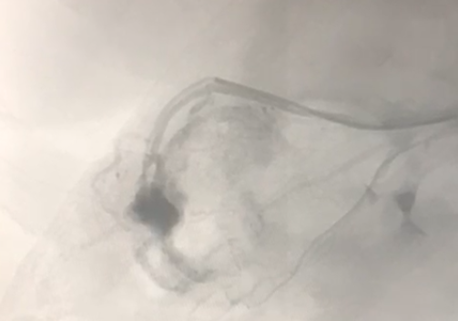
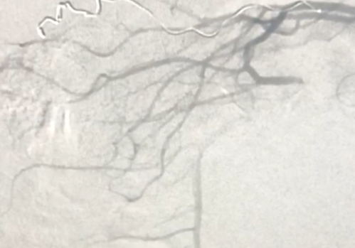
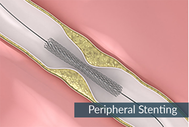
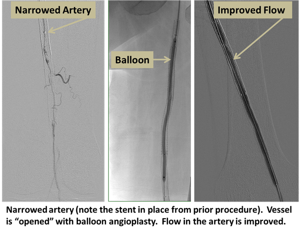
Narrowing of the arteries causing restricted blood flow (Peripheral Arterial Disease – PAD): Interventional radiologists treat this condition using balloons to widen the vessel (balloon angioplasty, PTA) and sometimes place metal mesh tubes called stents to keep the artery open. In some cases, arteries or bypass grafts can suddenly become blocked, leading to a rapid loss of blood supply to a limb. If blood flow is not restored, this may result in amputation. Interventional radiologists can help by delivering clot-dissolving medications directly into the artery through small catheters, often saving affected limbs. Dilated arteries (aneurysms) carry a risk of rupture and bleeding: Interventional radiologists treat these by lining the vessel with a tube called a stent graft.
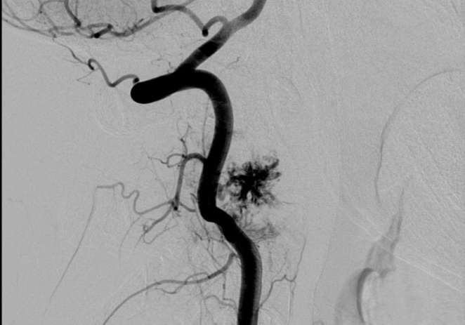
Bleeding (hemorrhage) is the most common vascular emergency treated by interventional radiologists. Hemorrhage can occur almost anywhere in the body, such as from the gastrointestinal tract, following major trauma, or after childbirth. Bleeding can often be permanently controlled by blocking the blood vessel (embolization), sealing it with a stent graft, or inflating a balloon within the vessel to stop the bleeding until emergency surgery can be performed. Interventional radiology is also used to help prevent bleeding during certain surgical procedures, such as cesarean sections in patients at high risk of hemorrhage due to abnormal placental conditions (postpartum hemorrhage).
Veins :
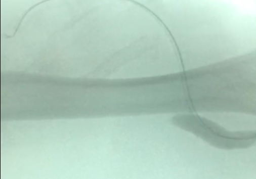
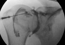
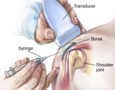
Blood clots in the lungs (Pulmonary Embolism, PE): Interventional radiologists use two main treatment approaches. They may place devices called inferior vena cava (IVC) filters to capture blood clots before they reach the lungs, helping to prevent further pulmonary embolisms. In cases of massive pulmonary embolism causing circulatory collapse, interventional radiologists can use small catheters to break up the clot and restore blood flow. Dilated veins (varicose veins): These are most commonly found in the legs but can also occur in the pelvis or scrotum. They can be treated by closing the affected vein using thermal techniques (laser or microwave ablation) or through the use of sclerosing agents and embolization techniques. Your doctor will first assess all affected veins using a duplex ultrasound examination and determine the best site for catheter insertion. You will be asked to wear protective eyewear if laser treatment is used. The treatment area will then be cleaned, shaved if necessary, and numbed with a local anesthetic.
Once the area is numb, a small incision is made, and a catheter along with a guidewire is inserted through the skin. A laser fiber is then advanced through the catheter until it extends approximately 1 to 2 centimeters beyond its tip, after which it is secured in place. The laser energy seals the faulty vein, and blood flow is naturally redirected to healthy veins. The entire procedure typically takes about one hour.
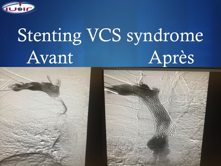
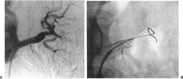
Blocked veins: This may occur as a result of a blood clot forming within a vein (Deep Vein Thrombosis, DVT), which can sometimes be treated by delivering clot-dissolving medications (thrombolysis) through a small catheter inserted into the vein. In some patients, blood clots develop due to a narrowing of the vein; once the clot has been dissolved, the narrowing can be treated using balloons and stents. In other cases, tumors within the chest may compress a vein, leading to facial swelling, headaches, and other symptoms that can often be relieved through the placement of a stent.
2. Non-Vascular Interventions
The Arteries:
Interventional oncology is often associated with cancer treatment, but these therapies are also effective for benign conditions. Interventional radiology treatments are used for the following purposes: to treat tumors or cancer (tumor ablation, embolization).
Tumor therapies: These treatments are designed to shrink or destroy tumors at their primary site or in areas where they have spread (metastases). This is a rapidly growing field of interest that can improve survival rates while reducing morbidity.
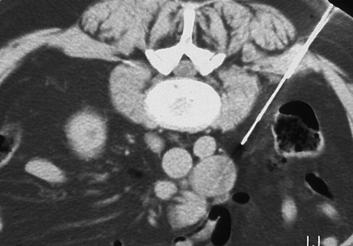
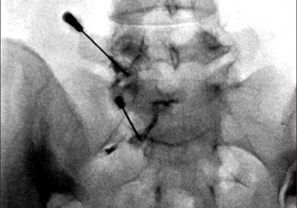
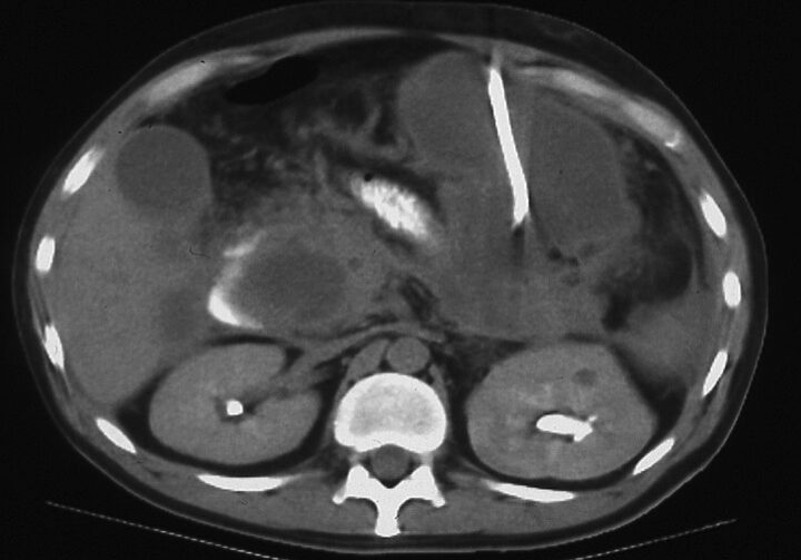
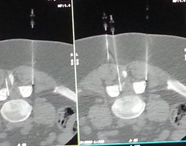
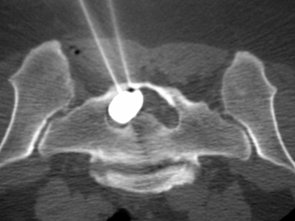
Liver tumors, kidney tumors, and other tumors (such as those affecting the bones or lungs) can be treated using ablative therapies that destroy tumor tissue, typically through heat-based techniques (radiofrequency, laser, microwave, or ultrasound ablation) or cold-based techniques (cryoablation). These treatments are performed and monitored using imaging guidance such as ultrasound, computed tomography (CT), or magnetic resonance imaging (MRI).
Uterine fibroids: Heavy menstrual bleeding and pelvic pain may be caused by benign tumors known as fibroids. These can be treated by blocking their blood supply (Uterine Fibroid Embolization, UFE), causing them to shrink over time. Embolization may also be combined with chemotherapy (chemoembolization) or radiation therapy (radioembolization), allowing treatment to be targeted directly at the tumor while reducing some of the side effects associated with cancer therapies.
3. Some of Our Procedures